
Translate this page into:
Rare case alert: Ochronotic arthropathy and its skeletal manifestations in 2 Indian siblings
Address for correspondence: Dr. Hemanth D Ramaiah, #8, Dr. Kuvempu Road, R S Palya, Kammanahalli Main Road, Bangalore 5600033. E-mail: hemanth.dr19@gmail.com
-
Received: ,
Accepted: ,
This article was originally published by Wolters Kluwer - Medknow and was migrated to Scientific Scholar after the change of Publisher.
How to cite this article: Kammar SF, Hosangadi AA, Ramaiah HD. Rare case alert: Ochronotic arthropathy and its skeletal manifestations in 2 Indian siblings. J Orthop Spine 2023;11:35-9.
Abstract
Alkaptonuria is a rare inborn error of metabolism disorder, with an incidence of one in a million births presenting with a triad of dark staining of urine, ochronosis, and Spondyloarthropathy. Till date, 1233 cases have been reported. Ochronotic arthropathy has often been a “serendipity,” with most diagnosis being made intraoperatively when the operating surgeons encountered “black” discs or “bluish-black cartilage. Spine involvement often precedes other musculoskeletal symptoms and begins after 30 years. Symptomatic treatment and arthroplasty form the mainstay of treatment as this disorder has no cure. We report a case of two brothers with a history of lumbar disc prolapse with radiculopathy, for which they underwent a surgery in their early thirties. They were asymptomatic for 6 years following the surgery before developing back stiffness and multiple large joint pains. The brothers were treated as HLA-B27 negative ankylosing spondylitis by multiple clinicians for 3 years before presenting to us with fused vertebrae, advanced arthritis of shoulder and knee, and avascular necrosis of bilateral femoral head. Alkaptonuria is a very rare disease; the symptoms of progressive low back ache/stiffness coupled with arthropathy of axial/weight bearing joints in an individual over 30 years should prompt the clinician to consider a diagnosis of alkaptonuri. Arthroplasty is the only solution to a pain-free functional life; however, it has its own complications. Hence, early diagnosis and lifestyle modifications might help delay the natural course of this debilitating disease until newer therapies emerge.
Keywords
Alkaptonuria
homogentisate oxidase deficiency
ochronosis
ochronotic arthropathy
spondyloarthropathy

Introduction
Alkaptonuria is a rare inborn error of metabolism, with a low global incidence as low as 0.001%–0.025% (higher incidence in Slovakian population, Dominican republic, and Jordan) linked to the deficiency of homogentisate 1,2 dioxygenase (HGO), an intermediate enzyme in the tyrosine catabolic pathway. It occurs as a result of loss of function mutation in the homogentisate deficiency gene on chromosome 3q21-q23 encoding for HGO enzyme leading to this disorder, which presents with classic triad of dark urine, ochronosis, and ochronotic arthropathy. Men are more commonly affected than women (2:1). Till date, 1233 cases have been reported, the earliest verified case of this disorder being the 3500-year-old mummy Harwa (a granary custodian who lived in 1500 BC). Though over the last century much progress has been made regarding the understanding of the disorder, we are yet to find a cure, and management is usually symptomatic. In this article, we report an occurrence of two cases of alkaptonuria among two brothers and perform a review of literature.[1,2,3,4]
Case Reports
Case I
A 44-year-old farmer walked in to the Orthopedic OPD; he had a cautious wide-based gait with a short stride and reduced hip and knee movements. He tells that he was apparently well until 31 years old, and when he developed sudden onset of low back ache following strenuous activity at farm and was managed as acute lumbosacral spasm, he recovered in 1 week with analgesics and rest. Patient was asymptomatic for the next year, and then, he developed a similar pain; however, this time over the course of 6 months, he developed tingling and numbness along right knee leg and ankle. straight led raising test was positive at 60° at the right lower limb, and the right Extensor Hallucis Longus power was 4/5. Magnetic resonance imaging showed multiple-level disc bulge in thoracic and lumbar with L4–L5 diffuse disc bulge with paracentral protrusion with nerve root compression for which he underwent a L4–L5 flavectomy + right foraminotomy in 2012. Following this, patient’s neurological symptoms improved and had relief of back pain; however, his stiffness worsened, and over the next 2 years, patient could not bend forward.
Six years following the spine surgery, he developed insidious onset pain in bilateral shoulder, hip, and knees, which over the course of next 2 years worsened such that he was unable to comb his hair or wear a T-shirt and was unable to sit cross legged. His walking distance had reduced from 5 km without pain following the spine surgery to 100 m with crippling pain over 6 years despite multiple visits to multiple specialists and was treated as HLA B27 negative ankylosing spondylitis. Since the past 2 years, he has developed this cautious gait and is unable to squat. On enquiring retrospectively, he gives a history of noticing his urine turn black since sixth grade, and because his elder brother had the same, he ignored it. His family history otherwise was unremarkable.
On examination, patient had small brownish black spots over bilateral sclera, bilateral tender shoulder joints with painful severe restriction of movements flexion 30°, extension 20°, abduction 30° predominantly scapula-thoracic, and internal and external rotation 5° and 15° bilaterally. Bilateral hip joints were tender (anterior joint line tenderness positive) with painful restricted internal rotation and positive sectoral sign with no fixed deformities. Bilateral knees showed medial joint line tenderness and flexion of about 80° (further flexion 10°) associated with crepitus throughout. Chest expansion was 4 cm, Schober’s test was 2.4 cm, and no Sacroiliac joint tenderness was seen. Cardiovascular, respiratory, per abdomen and neurological examination was within normal limits.
Plain radiographs demonstrated arthritic changes in bilateral shoulders with Grade II avascular necrosis changes in bilateral hips and bilateral knee Grade III Osteoarthritis. The sequential lumbosacral and thoracolumbar radiographs showed progressive reduction of intervertebral disc spaces with calcification with anterior osteophytes and end plate sclerosis [Figure 1].
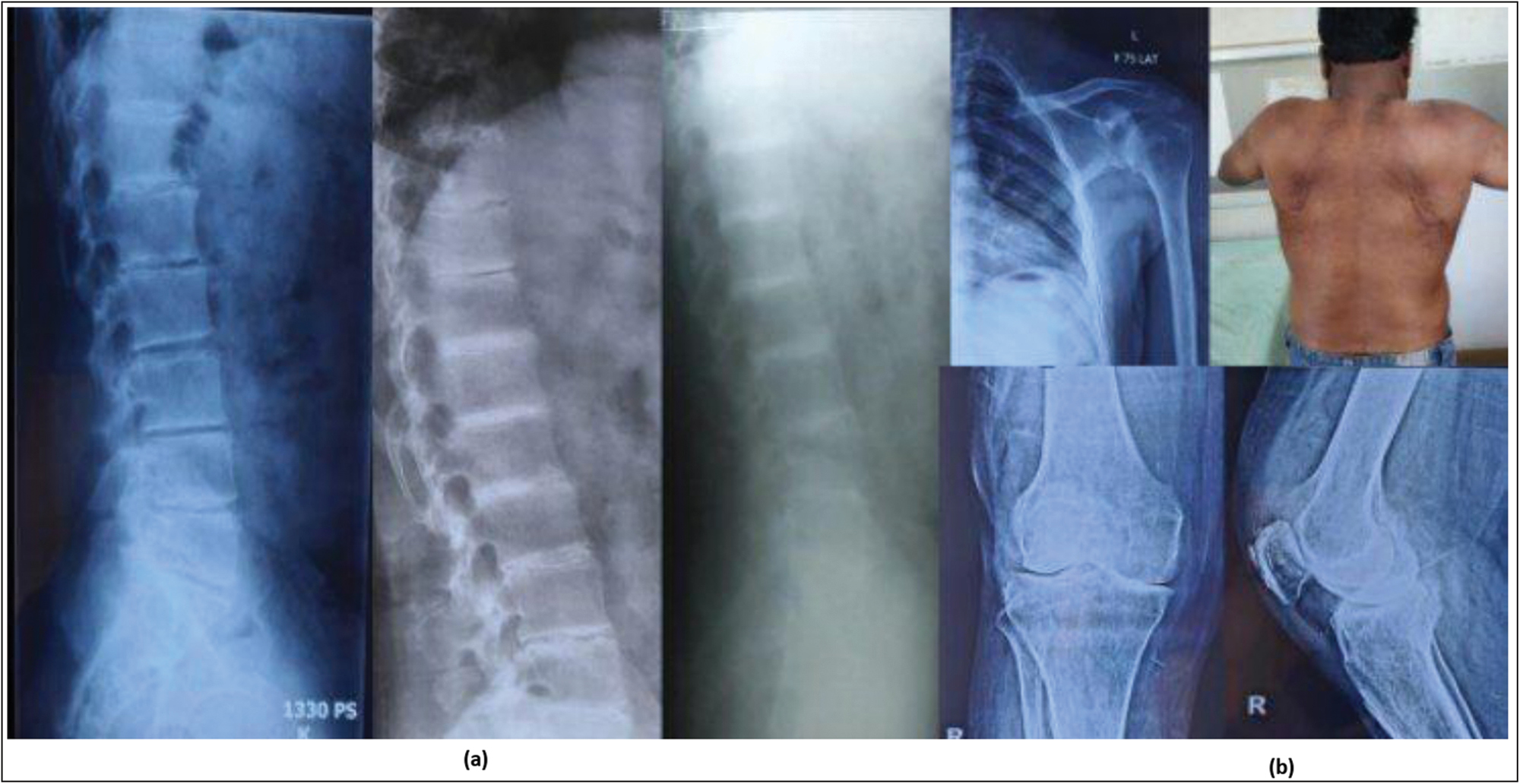
- Case I – (a) Serial TL spine Radiographs taken in 2011, 2017 and 2022 demonstrating progressive reduction of intervertebral disc space, marginal erosions and intervertebral disc calcifications. (b) Patient currently presented with severe restriction of abduction and other movements at shoulder joints in addition to arthritis of weight bearing joints and reduced spine flexion, classically noted in Ochronotic arthropathy
Case II
His elder brother, also a farmer, was 50 years old and had a similar course of disease with a L4–L5 discectomy in 2011 following which his back ache disappeared, and 5 years later, he developed similar symptoms of back stiffness, and hip and knee pain such that, since the past 1 year, he has been housebound. There was no history of alcohol or tobacco consumption in both patients. However, since the past 6 months, he has been taking 0.1-mg dexamethasone tablets prescribed by an AYUSH practioner in his village for joint pain.
On examination, his face was a little darker than the rest of his body, and he had black spots over left sclera. Bilateral hip examination revealed a flexion of 50° (further flexion 15°), internal and external rotations of 5° and 15° (with positive sectoral sign), and extension of 5° present at left hip. Harris hip score was 27. Knee joint examination showed bilateral tender joint line with a near normal range of movements, associated with crepitus. Shoulder joint examination was unremarkable. Chest expansion was 5 cm, and Schober’s test showed an increase of 1.8 cm against 5 cm. Other systemic examination was within normal limits.
Radiographs of pelvis with hips showed Grade IV avascular necrosis changes in both hips with visible cysts and subchondral sclerosis in bilateral femoral heads with acetabular involvement. Knee radiographs demonstrated advanced osteo-arthritis of bilateral knee joint. Computerized tomography (CT) scan of the lumbosacral spine demonstrated significant reduction in joint space with osteophytes, vacuum phenomenon, and Schmorl nodules [Figure 2].
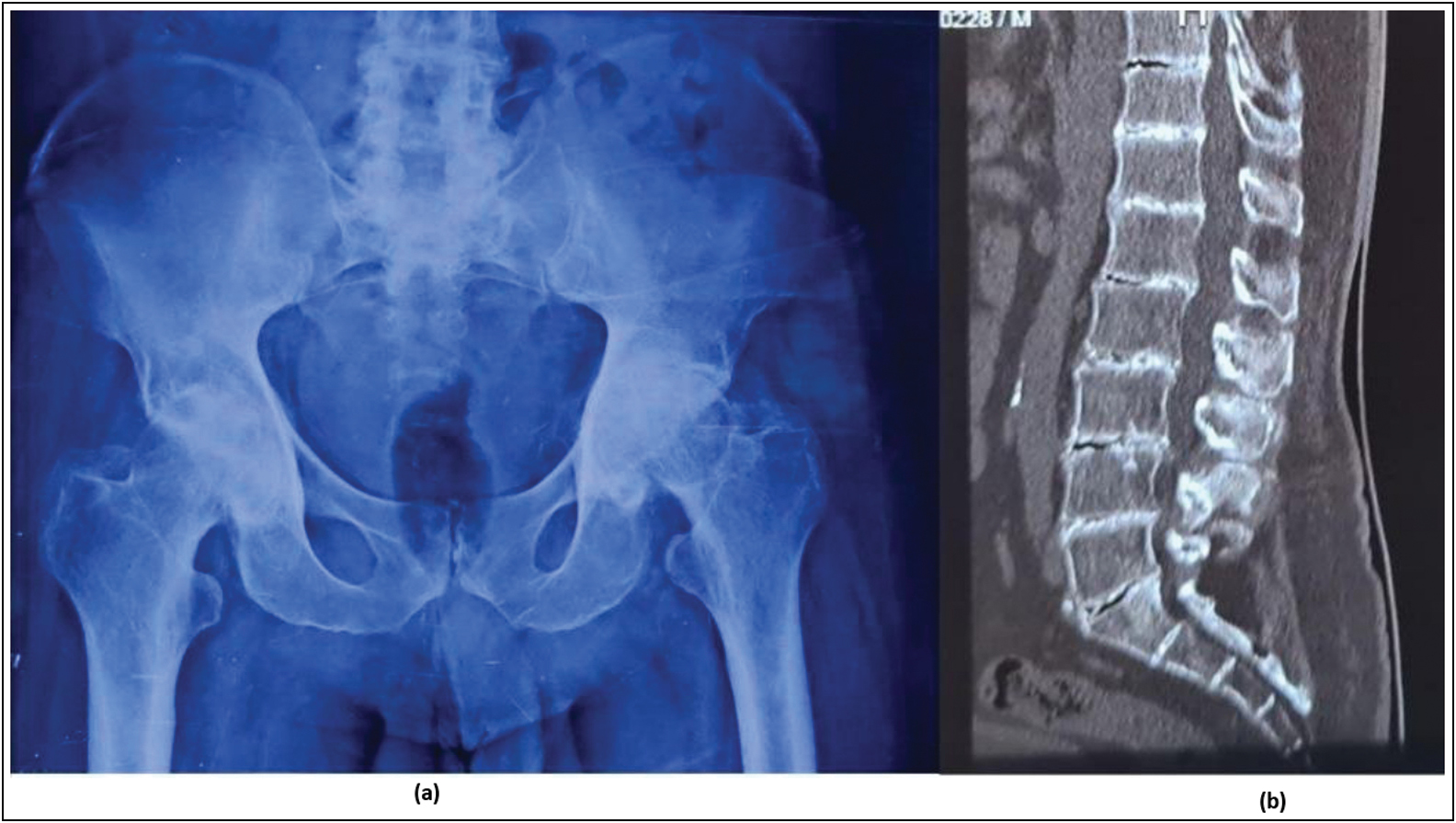
- Case II - (a) Plain radiograph of Pelvis with both hips demonstrating reduction in joint space of both hips with subchondral cysts in both femoral head and sclerosis of acetabular wall (Ficat and Arlet Stage IV). (b) CT scan of Lumbosacral spine shows Vacuum phenomenon (accumulation of air/gas in intervertebral disc spaces) and Schmorl's nodes indicating the degenerative nature of this disorder
Routine blood investigations of both patients were unremarkable, except ESR-45 mm/h. Ultrasonograph abdomen, electrocardiograph, and two-dimensional echocardiography showed normal rheumatoid arthritis factor, and C-reactive protein was negative. Urine analysis demonstrated a positive Benedict’s, ferric chloride, and ammoniacal silver nitrate tests [Figure 3]. Quantitative analysis of homogentisic acid (HGA) was 575.16 times normal. Patients were counseled about the nature of the disease and the possible treatment options; however, because of the sociocultural background, patient was skeptical about joint replacement at present. Patients chose to undergo a trial of physiotherapy for shoulder, spine, hip, and knee, with analgesics and low tyrosine diet and review in OPD when willing for surgery.
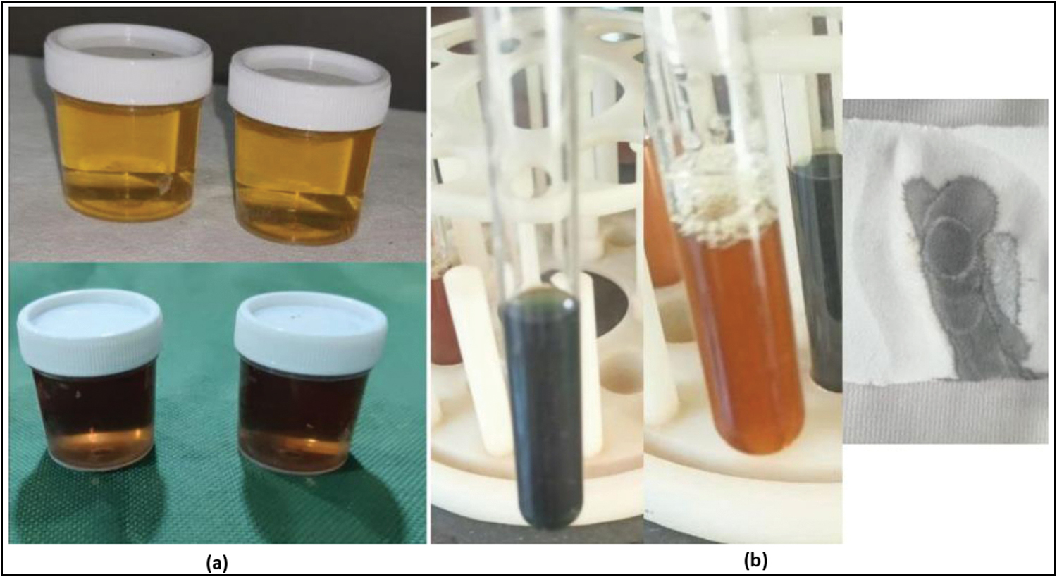
- Simple reliable tests to detect Alkaptonuria. (a) The most simple and economical test is the classical darkening of urine on exposure to air/sunlight due ton oxidation of Homogentisate and its by-products. (b) Tests for reducing products like Ferric Chloride, benedict's test and Ammoniacal silver nitrate was positive due to the characteristic reducing nature of the urine.
Discussion
The first description of alkaptonuri (AKU) in literature dates back to by Scribonius in 1584, when he mentioned a report of a school boy who passed urine as “black as ink.”[5] The term alkaptonuria originated from the Greek work “Alkapton,” which meant to suck up oxygen “greedily” in an alkali, based on Boedeker’s findings of reducing properties of patient’s urine. The term “Oochronosis” was coined by Virchow in 1866 in whom histologically described the connective tissue in AKU, given the cartilage’s “ochre” (yellow) hue under the microscope.[6] However, it is Sir Archibald Garrod, an English Physician (and also “the founding father of biochemical genetics”) who was credited with the discovery of the disease in 1902. He not only described all the features of AKU but also successfully mapped the inheritance of the disease by observing the diaper of a new-born whose older sibling and mother had already been diagnosed with AKU, and so, AKU became the first autosomal recessive disorder to be known to man. Further notable contributions were made by La Du in 1958, who showed absent HGO enzyme in the liver of patients affected with akaptonuria, and Pollak et al.[7] who identified the location of the homogentisate deficiency gene on chromosome 3q2.
The disease is first noticed in infancy when there is blackening of urine/diapers on standing, caused by oxidation of homogentisic acid to benzoquinone acetic acid and polymerization. This sign may never be noted; hence, the diagnosis is often delayed until adulthood. The declining renal clearance with progressing age leads to a steady level of high circulating levels in blood, and the unexcreted oxidized polymers bind to form macromolecules, which damage the collagen in cartilage present in joints, intervertebral discs, and other connective tissues like skin, ear, eye, tendons, and myocardial tissues and are responsible for the bluish black color.[8] Patients, therefore, typically present with a progressive degenerative arthropathy mainly affecting the axial and weight bearing joints with extraarticular manifestations usually after 30 years old.
The initial presenting symptom in majority of patients is back pain and stiffness (60% cases) and sciatica is initial presenting symptom in 17% cases.[9] Most of the cases are usually misdiagnosed as simple mechanical pain for years and eventually develop arthritis of the large peripheral joints like hip, knee, and shoulder over the next few years. Ochronotic arthropathy is primarily a degenerative peripheral arthropathy; however, at times, it presents with effusions and have a characteristic waxing and waning course like inflammatory arthropathies secondary to the body’s response to the benzoquinone polymers in the articular cartilage. This peaks by the fourth or fifth decade, and they usually warrant joint replacements by the age of 55 years.
Extraarticular manifestations include blackish pigmentation of palms, ear cartilage, face, sclera, spontaneous tendon ruptures, renal stones and obstructive uropathy, cardiac valvular calcifications, restricted lung disease, ventricular hypertrophy, and arterial intimal calcifications leading to myocardial infarctions. The average age at cardiac valve involvement, coronary artery calcifications, and renal stone formation is 54, 59, and 64 years, respectively. Hence, screening at regular intervals to look for these complications should be carried out from the fourth decade. Patients affected with alkaptonuria have a normal life expectancy, however a less functional one due to the severe arthritis.
Diagnosis is often delayed as the symptoms mimic ankylosing spondylitis and early osteoarthritis for years before the characteristic radiographic findings of calcified intervertebral discs appear with osteopenia of verterbral bodies. These dense calcifications occur in the nucleus pulposus as opposed to the annular calcifications seen in hemochromatosis, hypervitaminosis, hyperparathyroidism, DISH, pseudogout, aging, and even ankylosing spondyltitis. CT scan may reveal characteristic disc calcifications, syndesmophytes, vacuum phenomenon, and Schmorl nodules. Magnetic resonance imaging may show multiple-level disc bulges with T2 hypointensities.[10] All of the above were seen in our patients. Plain radiographs of the affected large joints may show symmetrical joint space narrowing with osteophyte formation, subchondral cysts and sclerosis, and extensive soft-tissue calcifications.
Ochronotic arthropathy should be differentiated from exogenous ochronosis, which occurs as a result of various drugs and chemicals like hydroxychloroquines, minocycline, methyldopa, phenol, and antimalarials.[11] Urine analysis for reducing substances and homogentisic acid levels may aid in differentiating between the two. The historic alkali test demonstrates darkening of urine on addition of caustic soda, ferric chloride test yields a characteristic green color, and test for ammoniacal silver nitrate is usually positive as indicated by blackish color. However, more reliable tests for diagnosis of alkaptonuria are identification of homogentisitic acid in urine by gas chromatography-mass spectrometry.[12]
There is currently no approved treatment for alkaptonuria. Various treatment modalities have been tried, earliest dating back to 1940s when Sealock et al studied the use of ascorbic acid for the treatment of alkaptonuria in rats.[13] It was found to delay the darkening of urine and also reduced the binding of HGA to connective tissues in vivo; however, it failed to replicate the same in humans or reduce the excretion of HGA in urine. For decades, symptomatic treatment of arthritis and specific treatment of extraarticular manifestations have been the mainstay of treatment in addition to dietary restriction of tyrosine and phenylalanine in diet. However, in the past decade, nitisinone, a synthetic reversible inhibitor of 4-hydroxy-phenylpyruvate-dioxygenase, has gained popularity in the treatment of alkaptonuria.[14] Clinical trials have demonstrated reduction in the levels of excretion of HGA in urine, decreased ochronosis, and showed improvement in symptoms. However, there have been reported side effects secondary to hypertyrosinemia such as dry eye, keratopathy, ocular hyperemia, and elevation of liver transaminases. However, this drug is still unavailable in most countries, and more clinical trials are needed in a diverse population group. Until then, dietary restriction of tyrosine, early onset physiotherapy, psychotherapy, and occupation change may improve the quality of life in these patients until they develop severe arthritis of joints. Replacement of joints such as hip, knees, and shoulder has been on the rise in recent times and has demonstrated a good clinical outcomes.[15]
Declaration of patient consent
The authors certify that they have obtained all appropriate patient consent forms. In the form, the patient(s) has/have given his/her/their consent for his/her/their images and other clinical information to be reported in the journal. The patients understand that their names and initials will not be published and due efforts will be made to conceal their identity, but anonymity cannot be guaranteed.
Financial support and sponsorship
Nil.
Conflicts of interest
There are no conflicts of interest.
References
- Alkaptonuria: A hereditary disease which is usually diagnosed in adulthood. Clin Dermatol Rev. 2021;5:200-3.
- [CrossRef] [Google Scholar]
- Musculoskeletal manifestations of alkaptonuria: A case report and literature review. Eur J Rheumatol. 2018;6:98-101.
- [Google Scholar]
- Total knee arthroplasty in ochronosis. Arthroplast Today. 2015;1:77-80.
- [CrossRef] [PubMed] [Google Scholar]
- Mutation spectrum of homogentisic acid oxidase (HGD) in alkaptonuria. Hum Mutat. 2009;30:1611-9.
- [CrossRef] [PubMed] [Google Scholar]
- Homozygosity mapping of the gene for alkaptonuria to chromosome 3q2. Nat Genet. 1993;5:201-4.
- [CrossRef] [PubMed] [Google Scholar]
- Analysis of melanin-like pigment synthesized from homogentisic acid, with or without tyrosine, and its implications in alkaptonuria. JIMD Rep. 2017;35:79-85.
- [CrossRef] [PubMed] [Google Scholar]
- Diagnosis of alkaptonuria after lumbar discectomy: Case report and a review of the literature. Fırat Tıp Dergisi. 2012;17:178-181.
- [Google Scholar]
- Alkaptonuric patient presenting with black disc and multiple Schmorl’s nodes. Int J Adv Res. 2016;4:1969-73.
- [CrossRef] [Google Scholar]
- Quick diagnosis of alkaptonuria by homogentisic acid determination in urine paper spots. JIMD Rep. 2017;31:51-56.
- [CrossRef] [PubMed] [Google Scholar]
- Administration of ascorbic acid to an alkaptonuric patient. Proc Soc Exp Biol Med. 1940;45:580-3.
- [CrossRef] [Google Scholar]
- Adequacy of nitisinone for the management of alkaptonuria. Ann Med Surg (Lond). 2022;80:104340.
- [CrossRef] [Google Scholar]
- Long-term outcomes of the knee and hip arthroplasties in patients with alkaptonuria. Arthroplast Today. 2020;6:689-93.
- [CrossRef] [PubMed] [Google Scholar]


