
Translate this page into:
High signal intervertebral disc in T1-weighted magnetic resonance imaging
Address for correspondence: Dr. Amit Agrawal, Department of Neurosurgery, Narayana Medical College Hospital, Chinthareddypalem, Nellore - 524. 003, Andhra Pradesh, India. E-mail: dramitagrawal@gmail.com
This article was originally published by Wolters Kluwer - Medknow and was migrated to Scientific Scholar after the change of Publisher.
Dear Sir,
A normal intervertebral disc is described as a homogeneous structure on magnetic resonance imaging (MRI), which is isointense to muscle on T1-weighted (T1W) and because of the water content becomes hyperintense on T2W images.[1,2,3] Most of the studies describe believe that because of immobile protons in calcium and complete maturity of calcium intervertebral disc calcification (IDC) appears signal void (dark) on T1W images (cortical bone).[4] Uncommonly IDC can be seen as high signal intensity on T1W images, probably due to the presence of mobile protons (liquid state or milk of calcium) in calcium.[4,5,6,7,8] A 60-year-oldgentlemen presented with the history of road traffic accident hit by four wheeler. There was no history of loss of consciousness; vomiting; ear, nose, and throat (ENT) bleed; or seizures. However, the patient was complaining of persistent neck which was made worse by neck movements. There was no radiation of pain. There were motor or sensory symptoms. Bowel and bladder functions were normal. There was no significant past medical history. The patient was examined with cervical spine radiograph and it showed fracture of anterior osteophyte at C2–3 and C3–4 level, bridging osteophytes, and hyperdensities in the intervertebral disc space at C4–5 level [Figure 1]. MRI of the cervical spine, T1W, T2W, and fluid-attenuated inversion recovery (FLAIR) images showed hyperintense signals in the C4–5 disc space with apparently normal spinal cord [Figure 2a-d]. Based on MRI imaging, hemorrhage in the disc space was suspected; however, as there was significant evidence of calcification on radiographs, the patient was further planned for CT of the cervical spine. CT cervical confirmed the presence of calcification in the C4–5 disc space [Figure 3a and b]. The patient was managed conservatively, he improved in his neck pain and doing well at follow-up.
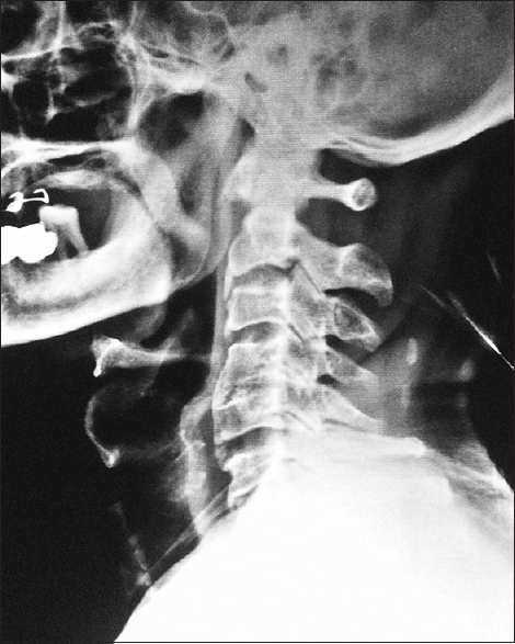
- Radiograph of the cervical spine lateral showing diffuse degenerative changes, fracture of the C2–3 and C5–6 anterior osteophyte, C4–5 bridging osteophytes, and intervertebral disc calcification
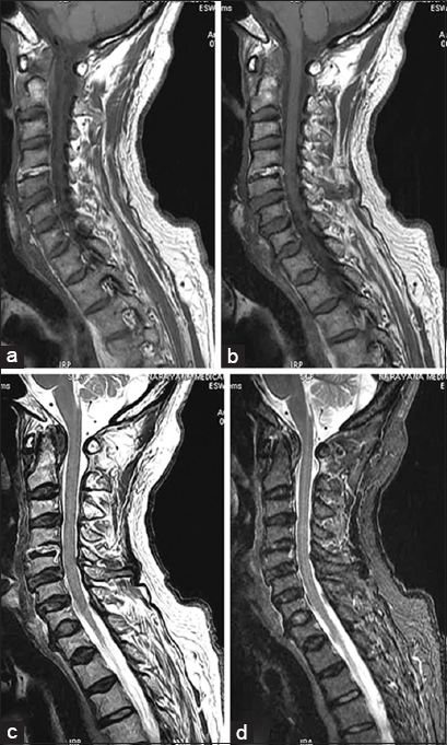
- Magnetic resonance imaging (MRI) cervical spine (a and b) T1-weighted (T1W), (c) T2W, and (d) fluid-attenuated inversion recovery (FLAIR) images showing hyperintense C4–5 intervertebral disc
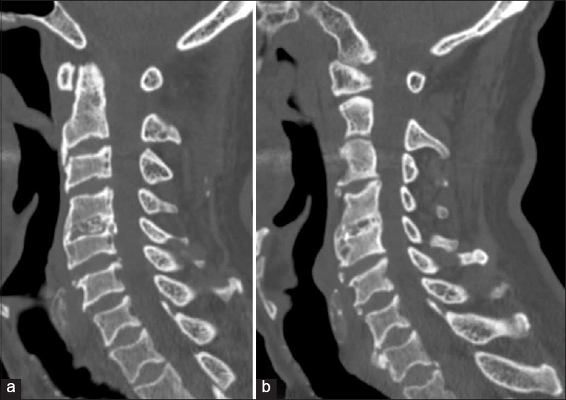
- Computed tomography (CT) scan cervical confirmed that the hyperintense appearance of intervertebral disc was due to extensive calcification
Chondrocalcinosis or IDC is characterized by the deposition of hydroxyapatite or calcium pyrophosphate dihydrate crystals within annulus fibrosus and/or nucleus pulposus of the intervertebral disc.[9] In a review it was noted that the IDC was described as early as 1,838 in anatomy textbooks and on radiographs in 1922.[10] On conventional radiography, the prevalence of IDC in the general adult population reported as 5–6% with[10,11] which had noticed higher in cadaver studies[12,13] and on histological examination of surgically retrieved disc materials.[14] IDC needs to be differentiated with many other disease conditions (e. g., hemochromatosis, chondrocalcinosis, hyperparathyroidism, poliomyelitis, acromegaly, amyloidosis, AIDS, multiple myeloma, myelofibrosis, Waldenstrom macroglobulinemia, and metastasis) which can lead to disc calcification and where the disc can appear hyperintense on T1W images.[10,11,15,16,17,18,19] Plain radiography has been recommended to identify intradiscal calcification.[2,20] The present case illustrates that hyperintense appearance of intervertebral disc on MRI can be misleading (particularly in presence of history of trauma) and need careful assessment and supportive investigations to differentiate hemorrhage from age-related changes particularly calcification.
REFERENCES
- Pitfalls, variants, and artifacts in body MR imaging. St. Louis, Missour: Mosby; 1996.
- [Google Scholar]
- Diffuse intervertebral disk calcification in a patient with rheumatoid arthritis. J Med Invest. 2000;47:152-4.
- [Google Scholar]
- Signal changes in the intervertebral discs on MRI of the thoracolumbar spine in ankylosing spondylitis. Clin Radiol. 1995;50:377-83.
- [CrossRef] [PubMed] [Google Scholar]
- Hyperintense disks on T1-weighted MR images: Correlation with calcification. Radiology. 1995;195:437-43.
- [CrossRef] [PubMed] [Google Scholar]
- Calcification demonstrated as high signal intensity on T1-weighted MR images of the disks of the lumbar spine. Radiology. 1993;189:494-6.
- [CrossRef] [PubMed] [Google Scholar]
- High signal intensity of intervertebral calcified disks on T1-weighted MR images resulting from fat content. Skeletal Radiol. 2005;34:80-6.
- [CrossRef] [PubMed] [Google Scholar]
- High signal intensity in MR images of calcified brain tissue. Radiology. 1991;179:199-206.
- [CrossRef] [PubMed] [Google Scholar]
- Intervertebral disc calcification in adults: A review. Semin Arthritis Rheum. 1978;8:69-75.
- [CrossRef] [PubMed] [Google Scholar]
- Intervertebral chondrocalcinosis: A coincidental finding possibly related to previous surgery. Arch Pathol Lab Med. 1980;104:269-71.
- [Google Scholar]
- Intervertebral disk calcification of the spine in an elderly population: Radiographic prevalence, location, and distribution and correlation with spinal degeneration. Radiology. 2004;230:499-503.
- [CrossRef] [PubMed] [Google Scholar]
- Radiological prevalence of lumbar intervertebral disc calcification in the elderly: An autopsy study. Skeletal Radiol. 1996;25:231-5.
- [CrossRef] [PubMed] [Google Scholar]
- Calcium pyrophosphate dihydrate deposition in lumbar disc fibrocartilage. J Rheumatol. 1981;8:955-8.
- [Google Scholar]
- Intervertebral disk calcification associated with spine fusion. Radiol. 1977;125:57-61.
- [CrossRef] [PubMed] [Google Scholar]
- Roentgenologic manifestations of juvenile rheumatoid arthritis. Am J Roentgenol Radium Ther Nucl Med. 1962;88:400-23.
- [Google Scholar]
- Waldenström macroglobulinemia: MR imaging of the spine and CT of the abdomen and pelvis. Radiology. 1993;188:669-73.
- [CrossRef] [PubMed] [Google Scholar]
- Magnetic resonance imaging of bone after radiation. Magn Reson Imaging. 1988;6:301-4.
- [Google Scholar]
- Fatty replacement of spinal bone marrow due to radiation: Demonstration by dual energy quantitative CT and MR imaging. J Comput Assist Tomogr. 1989;13:463-5.
- [CrossRef] [PubMed] [Google Scholar]
- Hypersignal of the intervertebral disks in T1-weighted spin-echo MRI sequences. J Radiol. 1994;75:363-7.
- [Google Scholar]


